S Ansar ![]() ,
H T AlGhosoon
,
H T AlGhosoon
For correspondence:- S Ansar Email: sansar@ksu.edu.sa Tel:+966118052970
Received: 8 September 2015 Accepted: 18 December 2015 Published: 29 January 2016
Citation: Ansar S, AlGhosoon HT. Effect of diallylsulphide supplementation on Wistar rats exposed to mercuric chloride. Trop J Pharm Res 2016; 15(1):81-86 doi: 10.4314/tjpr.v15i1.11
© 2016 The authors.
This is an Open Access article that uses a funding model which does not charge readers or their institutions for access and distributed under the terms of the Creative Commons Attribution License (http://creativecommons.org/licenses/by/4.0) and the Budapest Open Access Initiative (http://www.budapestopenaccessinitiative.org/read), which permit unrestricted use, distribution, and reproduction in any medium, provided the original work is properly credited..
Purpose: To evaluate the protective potential of diallylsulphide (DAS) against mercury-induced oxidative stress and antioxidant enzymatic alterations in spleen of rats.
Methods: Rats were randomly divided into 4 groups of 6 rats each and exposed to mercuric chloride (HgCl2) (50 mg/kg, i.p.) and/or diallylsulphide (200 mg/kg/b.w) by gavage. Oxidative stress was evaluated in spleen by antioxidant markers, viz, lipid per oxidation (LPO), superoxide dismutase (SOD), reduced glutathione (GSH), glutathione peroxidase (GPx) and catalase (CAT). Histomorphological changes in the spleen of rats were also compared between groups.
Results: Oral administration of DAS at a concentration of 50 mg/kg daily showed a significant increase in GSH and GPx (p < 0.05), SOD and CAT (p < 0.05), as well as decreased LPO (p < 0.05) level in the spleen of rats as compared with HgCl2 treated rats. Histopathology of spleen also showed that administration of DAS reduced the damage generated by HgCl2 treatment.
Conclusion: The results suggest that DAS may effectively normalize impaired antioxidant status in HgCl2-induced oxidative stress. DAS has a protective effect against lipid peroxidation by scavenging free radicals and is thus capable of ameliorating mercury-induced changes in the spleen of adult Wistar rats.
Introduction
Mercury toxicity has led to many health problems in the world. Mercury results in a variety of adverse neurological, renal, respiratory, immune, dermatological, reproductive and developmental disorders [1]. Its wide industry-related effects on humans have been well documented [2]. Mercuric chloride from industries, when released into water, causes morbidity and mortality in humans consuming sea food, including fish [3,4].
Exposure to mercury contamination could be from water, soil and air and can disturb the metabolic pathways [5]. It has been shown that kidneys contain the largest residues of mercury, followed by liver, spleen, intestine, lymph node, skeletal muscles, lungs, heart, brain, and the intensity of the cytotoxic changes in the various organs was proportional to the amount of mercury accumulated [6].
The spleen is a secondary lymphoid organ present in all vertebrates in which the parenchyma is divided into white pulp (WP) and red pulp (RP) distinguished by different colors. The WP contains dense and highly organized accumulations of B and T lymphocytes around arterioles, while most of the RP consists of blood‑filled spaces. These RP spaces are composed of two structures, the arterioles and veins forming the splenic sinuses [7]. Earlier it has been reported that sub-chronic exposure to mercuric chloride resulted in a transient decrease of the lymphoid cell ratio which affected the incidence of splenic T‑cell subsets [7-9].
The protective effects of garlic, and its constituents, have been attributed to presence of organosulphur compounds like diallyl sulphide (DAS), diallyl disulphide (DADS), ajoene, allixin, allyl mercaptans and allyl methylsulphides [10-12]. The DADS is one of the major volatile degradative compounds of garlic. Many studies on animals showed its protective effects against chemically induced toxicity and carcinogenesis [13,14].
Therefore, the present study was designed to study the antioxidant and protective effects of DAS against HgCl2 toxicity.
Methods
Animal groups
Male Wistar rats weighing approximately 180 - 200 g were used. All the animals used in this study were placed in cages in an air conditioned room maintained 12 h light/dark cycle. The study was approved by the Ethical Committee of King Saud University (CAMS 28-36/7) and was carried out in compliance with the declarations of National Research Council [15].
Experimental protocol
Thirty male Wistar rats were randomly divided into 5 groups (6 rats in each group). Group I received saline injection intraperitoneally (0.85 % NaCl) at a dose of 10 ml/kg body weight. Group II received a single intraperitoneal injection of mercuric chloride at a dose of 50 mg/kg bodyweight. Group III and IV received pretreatment with DAS i.p. once a day for 7 days at a dose of 200 mg/kg body weight. After the last treatment with DAS, the rats of group IV received a single intraperitoneal injection of HgCl2 at a dose level of 50 mg/kg body weight. The single doses of DAS and mercury selected were based on a published dosage regimen that has been shown to be biologically and therapeutically active in experimental systems. After 24 h of last administration, the animals were sacrificed by cervical dislocation and the spleen tissue was harvested for biochemical and histopathological studies.
The spleen of rats were washed in ice-cold 50 mM Tris–HCl, pH 7.4 and homogenized in ice-cold medium that contained 50 mM Tris–HCl, pH 7.4. The homogenates were centrifuged at 3000 rpm for 10 min at 4 °C. The supernatants were used for the various biochemical determinations. The total protein content of the homogenized spleen was determined using bovine serum albumin as a standard [16].
Assay of oxidative stress markers
MDA content was assayed using the thiobarbituric acid (TBA) test as described by Ohkawa [17]. After incubation of homogenate with TBA at 95 °C, MDA reacts to form a pink colored complex. Absorbance was measured spectrofotometrically at 532 nm to determine the MDA content. The specific activity is expressed as nmol/mg protein.
For measurement of glutathione (GSH), an aliquot of 1.0 ml of renal PMS (10 % w/v) was precipitated with 1.0 ml of sulphosalicylic acid (4 % w/v). The samples were kept at 4 oC for at least 1 h and then subjected to centrifugation at 1200 g for 15 min at 4 oC. The assay mixture contained 0.1 ml filtered aliquot, 2.7 ml phosphate buffer (0.1 M, pH 7.4) and 0.2 ml DTNB (40 mg/10 ml of phosphate buffer 0.1 M, pH 7.4) in a total volume of 3.0 ml. The yellow color developed was read immediately at 412 nm on a spectrophotometer. The amount of glutathione was expressed as mmol/g tissue [18].
Total SOD activity was determined according to the method described by Marklund and Marklund [19] by assaying the autooxidation and illumination of pyrogallolat 440 nm for 3 min. One unit of SOD activity was calculated as the amount of protein that caused 50 % pyrogallol autooxidation inhibition. The SOD activity is expressed as IU/mg protein.
Before determination of the CAT activity, homogenates were diluted 1:9 with 1 % (v/v) Triton X-100. CAT activity was measured according to the method described by Aebi [20] by assaying the hydrolysis of H2O2 and the resulting decrease in absorbance at 240 nm over a 3 min period at 25 oC. CAT activity is expressed as IU/mg protein.
GPx activity was measured using H2O2 as substrate according to the method described by Paglia and Valentine [21]. The reaction was monitored indirectly as the oxidation rate of NADPH at 240 nm for 3 min. Enzyme activity was expressed as U/mg protein.
Histological examinations
Spleen tissues from rats were fixed in 10 % formaldehyde. Fixed tissues were dehydrated in serial ethanol series, trimmed, embedded in paraffin, sectioned into 2-μm sections and stained with hematoxylin and eosin (H&E). Morphological examination was conducted under a light microscope (Nikon Eclipse E600).
Statistical analysis
The results are expressed as mean ± standard error of the mean (SEM). One-way ANOVA was applied to test for the significance of the biochemical data for the different groups. Significance level was set at p < 0.05.
Results
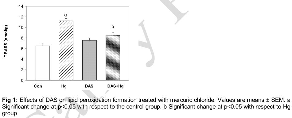
HgCl2 administration caused a significant (p < 0.05) increase in the level of LPO in brain compared to the control group. DAS pretreated group caused a significant (p < 0.05) reduction in LPO levels increased by HgCl2 administration as compared to control as shown in .
The HgCl2-induced oxidative stress was indicated by a significant reduction (p < 0.05) in GSH contents of spleen tissue of HgCl2-treated rats in comparison with the control group. This reduction in GSH contents was attenuated by pretreatment with DAS as shown in .
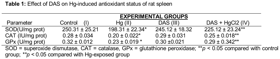
HgCl2 administration led to modulation of antioxidant enzymes relative to the control rats. It also led to decrease in SOD, CAT, and GPx activities in spleen homogenates significantly (p < 0.05) compared to the control (). On the other hand DAS pretreatment elevated the activities of SOD, CAT, and GPx significantly (p < 0.05) compared to the HgCl2 group.
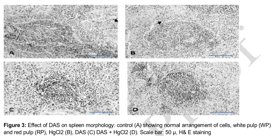
Histopathological examination of spleen showed changes in the rats administered with mercury chloride with or without DAS as shown in . The result from the control (A) showed normal spleen tissue showed normal arrangement of cells, white pulp (WP) and red pulp (RP). While the spleen of the rats in the HgCl2 treated group (B) showed changes in the spleen characterized by swelling, and monocular infiltration. While DAS pretreated group (C) and group (D) showed normalcy to architecture of the spleen which are similar to the control group (A).
Discussion
The present study shows the ameliorative effects of DAS on the spleen of experimental rats treated with mercury chloride. It is well known that heavy metals are widely distributed in the environment and some of them can cause physiological, biochemical and histological disorders. The degree of toxic manifestation of different metals depend on dose, duration, route of administration and other physiological factors has been shown in many past studies [22-24].
The mercury usually deposits in the liver, kidneys, nerve cells, heart and other skeletal muscles, whereas in chronic condition its deposition is observed predominantly in hard tissues. In this study, DAS has been shown to ameliorate the toxic effects of mercury chloride on the biochemical parameters and plays important role in preventing oxidative damage to the spleen. Histological examination revealed that in the mercury treated group, spleen showed increased infiltration and vacuolization of the cells. Non treated spleen sections showed normal spleen structure composed of white pulp and red pulp, besides fibrous capsule which covered the spleen. White pulp consists mainly of B lymphocytes arranged into two zones, marginal zone (outer rim of loose lymphocytes) that contains macrophages and mantle zone (inner rim of lymphocytes).
In rats treated with mercuric chloride, there was significant decrease in the levels of GSH and SOD in spleen tissue. Glutathione, a tripeptide present in the majority of cells, is responsible for hydrophilic xenobiotics conjugation. GSH serves many vital physiological functions including protection of cells from reactive oxygen species (ROS), detoxification of exogenous compounds, and amino acid transport. Sulphydryl group of glutathione is essential for its antioxidant activity against some forms of ROS in cells. Much of the pathology is associated with the decrease in intracellular GSH concentration. Therefore, GSH concentration is important for survival of the cells. MDA is the most abundant individual aldehyde resulting from lipid peroxidation breakdown in biological systems and is commonly used as an indirect index of lipid peroxidation. In this study, lipid peroxidation levels increased by HgCl2 administration as compared to control and DAS pretreated group caused a significant reduction as compared to mercury treated group.
The most important protective mechanism for free radical scavenging and superoxide dismutases (SODs) belongs to a family of antioxidant enzymes that catalyze the dismutation of superoxide to yield hydrogen peroxide and oxygen. SOD is essentially a protective enzyme which scavenges the superoxide ions produced as cellular by-products during oxidative stress. Its decreased activity can lead to adverse effects because superoxide anions are extremely toxic and may accumulate in the cells.
Supplementation of DAS to the mercuric chloride-treated groups ameliorated malondialdehyde, glutathione and antioxidant enzymes activities. This could be explained by the major role of natural antioxidants in inhibiting lipid peroxidation and in protecting the wholeness and functioning of tissues and cells. Antioxidants may play an important role in abating some health hazards of heavy metals in connection with an interaction of physiological free radicals. It has also been known that garlic and its related compounds have inhibitory effects on chemical carcinogenesis and mutagenesis. These compounds may inhibit the activation of other carcinogens at low efficiency and the induction of glutathione -S-transferase and phase II enzymes may also play a role. Henceforth, it could be interpreted that the tissue protection against mercury may be due to the free radical scavenger effect of DAS.
Conclusion
The findings of this study suggest that mercuric chloride intoxication causes reactive oxygen species generation which in turn induces biochemical and histological alterations in rats. Administration of DAS proved to be beneficial in attenuating the mercuric chloride-induced oxidative toxicity.
Declarations
Acknowledgement
References
Archives


News Updates